In the process of cell culture, there will be such problems. From the perspective of cell growth, we should do the following analysis and propose a corresponding solution for the reasons that cells are not adherent, slow growth, poor growth, or even death. method.
First, the culture cells are not attached
Possible Causes:
• excessive trypsin digestion;
• Mycoplasma contamination;
• The pH of the medium is over-alkali (NaHCO 3 decomposition);
• cell aging;
• The initial concentration of the inoculated cells is too low or too high.
Solution:
• shorten trypsin digestion time or decrease trypsin concentration;
• Isolation of cultures to detect mycoplasma. Clean the stand or incubator. If the mycoplasma is found to be contaminated, discard the culture;
• Use a sterile acetic acid solution to adjust the pH or fill with sterile CO 2 ;
• Enable new seed cells;
• Adjust the optimal inoculated cell concentration.
Second, the culture cells grow slowly
Possible Causes:
• due to the replacement of different culture fluids or serum;
• Some cells in the culture medium, such as glutamine or growth factors, are depleted or lacking or have been destroyed;
• There is a small amount of bacterial or fungal contamination in the culture;
• Improper storage of reagents;
• The initial concentration of the inoculated cells is too low;
• The cells are aging;
• Mycoplasma contamination.
Solution:
• Compare the new culture medium with the original culture solution, compare the new serum with the old serum to support the cell growth experiment, and let the cells gradually adapt to the new culture solution;
• Swap in freshly prepared culture medium or add glutamine and growth factors;
• Culture with antibiotic-free culture medium, discard the culture if contamination is found;
• Serum should be stored at -10 to -20 °C. The culture solution should be stored at 2-8 ° C in the dark. The serum-containing complete culture solution is stored at 2-8 ° C and needs to be used within 1 week;
• increase the initial concentration of seeded cells;
• Switch to new seed cells;
• Isolation of cultures to detect mycoplasma. Clean the stand and incubator. If the mycoplasma is found to be contaminated, discard the culture.
Third, the culture cells are not growing well.
Possible Causes:
• the state of the cell itself
Cell passage times are numerous, and cells are aged;
Inoculation amount of cells: the inoculum amount is too low, and the cell growth is slow;
Cell passage time is too late: cell poisoning, affecting cell growth after passage;
The trypsin digestion time is too long or too short: the time is too long, the cell is dead; the time is too short, the cells are not completely separated into a mass, and the cell is dead;
Cryopreservation and resuscitation of cells: slow freezing and rapid melting.
• Pollution
Mycoplasma contamination;
Mold contamination.
• Medium or serum
No verification was made prior to serum or media replacement;
Whether the selected medium is suitable;
Whether the medium is prepared accurately.
• Develop an environment
Whether the CO 2 supply is normal;
The incubator or shaker temperature control is correct.
Solution:
Make targeted solutions based on the possible reasons for the above four aspects
• Pay attention to the state of the cells themselves: such as the number of passages, the amount of inoculation, etc.;
• Avoid contamination (using regular, legal, traceable serum);
• Use appropriate serum or medium, preferably verified;
• Pay attention to the laboratory environment.
Fourth, culture cell death
Possible Causes:
• There is no CO 2 in the incubator;
• The temperature inside the incubator fluctuates too much;
• damage during cell cryopreservation or resuscitation;
• The osmotic pressure of the culture fluid is incorrect;
• The accumulation of toxic metabolites in the culture solution.
Solution:
• Detect CO 2 in the incubator;
• Check the temperature inside the incubator;
• Take new preserved cell types;
• Detect the osmotic pressure of the culture fluid;
• Change to fresh medium.
Here are the recommended Esco products - CelCradle series bioreactor - the best cradle for high-density cell culture
1. What is the CelCradle TM Bioreactor?
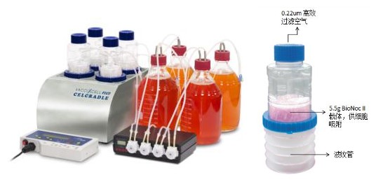
CelCradle TM bioreactor design made in accordance with the principles of the tides, wherein the compression and decompression of the bellows allows cells to nutrient medium constant contact with the air, provides a low cell tear, no foam, a high oxygen saturation and high Nutrient concentration of the cell culture environment.
CelCradle TM is a desktop bioreactor economical and easy to use a high density cell culture, cultured cells may be high density, cell secreted products may be collected.
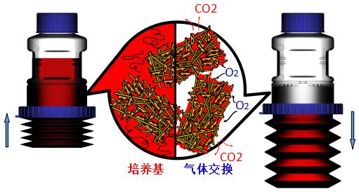
Second, CelCradle TM tidal working principle
Bottom of the culture flask CelCradle TM cells by compression and decompression of the bellows, the cell intermittently exposed to air and the medium, the cell culture medium and full contact with oxygen, and principles is similar to roller bottles.
Third, CelCradle TM bioreactor product features
• Large area high density adherent cell culture space
• Pre-sterilized, ready to use
• One-time use of CelCradle TM flasks for easy operation
• Low shear, low cell tear, no foam and no oxygen-limited high nutrient cell culture environment
• Compatible with most serum-free media
• Culture and collection of whole cells, cell components or cell products
Why choose the CelCradle TM bioreactor
1. Easy to use design, one-time use can save space and human resources
CelCradle TM bioreactor is a disposable bioreactor system is a readily available, no start-up time and complexity of the learning process.
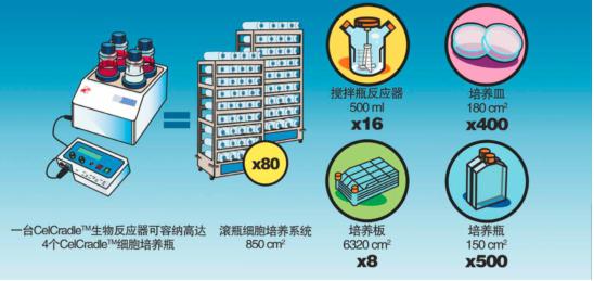
CelCradle TM bioreactor may be placed in a CO 2 incubator 6 cubic feet, and can operate four cell culture flasks simultaneously. A standard cell culture bottles CelCradle TM 5.5g BioNoc II with a carrier, provides 15,000 cm2 cell culture total area of cultured cells can reach 4 ~ 5x10 9 / vial, equivalent to 18 to 20 cell culture flask of rolling The amount of cultivation.
CelCradle TM culture by increasing the number of bottles or use of cell expansion of production scale to achieve the same principle as the tide of TideCell TM bioreactor, thus simplifying the process of scale culture systems and the savings required during development expenditures.
2. High cell yield, special B_H function promotes protein expression
A bioreactor CelCradle TM substitutable hundreds dish, and the traditional cell culture flask plurality of sets of cell culture roller bottles, etc., thus greatly reducing the production cost.
Not only does this system achieve high cell yields, but this special B_H function (increased bottom hold time) not only limits cell overgrowth but also increases protein expression by more than 3 fold while achieving higher protein concentrations. And save the medium during the cultivation process.
V. Application fields
• Mammal and insect cell culture
• Protein and virus production
• Monoclonal antibody production
• Proteome research
• Drug development
• Pharmacokinetic studies
• Gene and cell therapy research
Professional paper support
The following are some of the existing academic research applications support CelCradle TM cell culture system
[1]Akiyama, M., Nakayama, D., Takeda, S., Kokame, K., Takagi, J., & Miyata, T. (nd). Crystal structure and enzymatic activity of an ADAMTS-13 mutant with the East Asian-specific P475S polymorphism. J Thromb Haemost Journal of Thrombosis and Haemostasis, 1399-1406.
[2] Asaoka, Y., Tanaka, T., Tsumoto, K., Tomita, M., & Ide, T. (nd). Efficient expression of recombinant soluble human FcγRI in mammalian cells and its characterization. Protein Expression and Purification , 155-161.
[3] Brown, A., Mcsharry, J., Adams, J., Kulawy, R., Barnard, R., Newhard, W., . . . Drusano, G. (2011). Pharmacodynamic Analysis of a Serine Protease Inhibitor, MK-4519, against Hepatitis C Virus Using a Novel In Vitro Pharmacodynamic System. Antimicrobial Agents and Chemotherapy, 1170-1181.
[4]Chen, Y., Wu, J., Wang, K., Chiang, Y., Lai, C., Chung, Y., & Hu, Y. (nd). Baculovirus-mediated production of HDV-like Particles in BHK cells using a novel oscillating bioreactor. Journal of Biotechnology, 135-147.
[5]Drugmand, J., J.-F., J., Agathos, S., & Schneider, Y. (nd). Growth of Mammalian and Lepidopteran Cells on BioNOC ® II Disks, a Novel Macroporous Microcarrier. Cell Technology For Cell Products, 781-784.
[6]Hammonds, J., Chen, X., Zhang, X., Lee, F., & Spearman, P. (nd). Advances in methods for the production, purification, and characterization of HIV-1 Gag–Env Pseudovirion vaccines. Vaccine, 8036-8048.
[7]Haredy, A., Takenaka, N., Yamada, H., Sakoda, Y., Okamatsu, M., Yamamoto, N., . . . Okamoto, S. (2013). An MDCK Cell Culture-Derived Formalin-Inactivated Influenza Virus Whole-Virion Vaccine from an Influenza Virus Library Confers Cross-Protective Immunity by Intranasal Administration in Mice. Clinical and Vaccine Immunology, 998-1007.
[8] Haredy, A., Yamada, H., Sakoda, Y., Okamatsu, M., Yamamoto, N., Omasa, T., . . . Yamanishi, K. (2014). Neuraminidase gene homology contributes to the Protective activity of influenza vaccines prepared from the influenza virus library. Journal of General Virology, 2365-2371.
[9] Ho, L., Greene, C., Schmidt, A., & Huang, L. (nd). Cultivation of HEK 293 cell line and production of a member of the superfamily of G-protein coupled receptors for drug discovery Applications using a highly efficient novel bioreactor. Cytotechnology, 117-123.
[10] Hu, Y., Lu, J., & Chung, Y. (nd). High-density cultivation of insect cells and production of recombinant baculovirus using a novel oscillating bioreactor. Cytotechnology, 145-153.
Customers interested in learning more about our CelCradle TM series of bioreactors can contact us through the following contact methods:
Mobile: 010-5823 6368/5823 6468
Email:
Website:
This article was reprinted from CellMax fetal bovine serum, and has been adapted and typeset. If you have any objection, please contact us.
Suction blood vessel is also called vacuum collection blood vessel, according to the type of a total of nine categories. The details are as follows :1. Red head cover, no additives, used for the determination of some biochemical and immune indicators. 2. Yellow head cover, containing coagulant, can be used for biochemical and drug test determination. 3. Black cap, which is mainly used for the determination of erythrocyte sedimentation rate. 4. Light blue cap for the determination of coagulation factors. 5. Green cap for blood gas analysis and hematocrit measurement. 6. Light green head cover, liver function, blood lipid, blood sugar and so on were measured. 7. Grey cap, which is a special test tube for measuring blood sugar. 8. Purple head cover, mostly used for determination, blood type, blood routine, glycosylated hemoglobin and so on. 9 orange head cover, used for the determination of serum, hormones and so on.
Type of blood vessel extraction:
1, red head cover, no additives, used for the determination of some biochemical and immune indicators.
2, yellow head cap contains coagulant, can be used for the determination of biochemical and drug tests.
3. Black cap, which is mostly used for determining erythrocyte sedimentation rate.
4. Light blue cap for the measurement of blood coagulation factor.
5, green head cap, for gas and blood analysis, hematocrit measurement.
6, light green head cover, liver function, blood lipid, blood sugar and so on.
7. Grey cap. This is a special test tube for measuring blood sugar.
8, purple head cover is used for determination, blood type, blood routine, glycosylated hemoglobin and so on.
9, orange head cover, mostly used for serum, hormone and other measurements.
Blood Collection Tube,Red Glass Plain Tube,Red Pet Plain Tube,Gel&Clot Activator Tube,Yellow Color Blood Tube
Changchun ZYF science and technology CO.,LTD , https://www.zyf-medical.com