Release date: 2017-03-15
You may not see the poor life, how the cancer cells in the split grow, and those who have not heard of their names, but are really close to you, located in a tiny part of your body, they are also different. The beauty.
The Wellcome Image Awards recognizes the world of hidden worlds and shows the best science-related images of the past year. The 2017 finalists have announced that the final winners will be announced at the Wellcome Trust in London on March 15th. Open the mysterious journey of life.
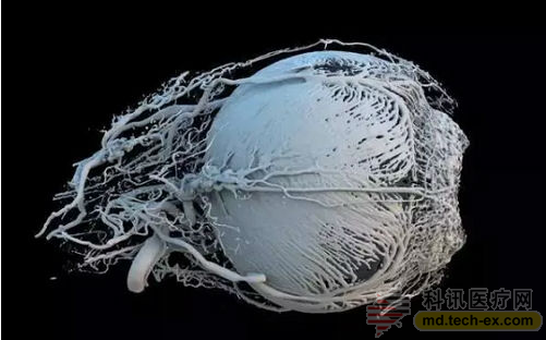
Researchers from Switzerland use computed tomography (CT) and 3D printing to present blood vessels in pig eyes in this unique way. The dense blood vessels provide energy and food to the muscles around the iris, which determines how much light can enter the eye. The pupil is on the far right.
Language Pathways of the Brain
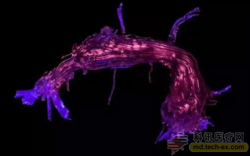
Also applied to 3D printing technology, this part is the white matter connection area between languages. The model is created by beam imaging technology, which uses a scanner to track the movement of water molecules in the brain.
Surface of a Mouse Retina (surface of mouse retina)
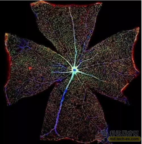
By stitching together more than 400 Microscope images, you can see a view of the mouse retina. The blue line in the figure is the blood vessels, and the astrocytes (special cells of the nervous system) are shown in red and green. By studying the functional changes of astrocytes during retinal degeneration, scientists hope to find new ways to treat vision impairment.
The Placenta Rainbow (Placenta Rainbow)
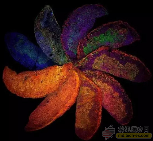
This is from a genetically modified mouse placenta, each with its own unique immune system. The production of "placental rainbow" also highlights differences in placental development, which can be controlled by the maternal immune system. Researchers can thus better understand and treat complications that occur during pregnancy in humans.
Unravelled DNA in a Human Lung Cell (loose DNA in human lung cells)
A new human lung cell nucleus filled with DNA. Cellular deformation is caused by the tension of the junction, which is produced by the linear DNA stretched between the two cells.
Developing Spinal Cord
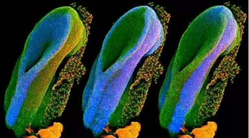
The spinal cord is composed of the tissue structure of the neural tube and is often formed during the first month of pregnancy. These three images show the open end of the rat's neural tube, and the blue portion of each image highlights a major embryonic tissue type. The one on the left will eventually form the brain, spine and nerves, the middle will grow into organs, and the right will eventually form skin, teeth and hair.
Zebrafish Eye (zebrafish eye)
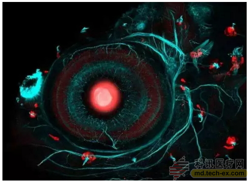
The picture above shows the eyes of a zebrafish embryo born four days ago. The zebrafish was nurtured by researchers at the University of London, and the researchers used the CRISPR-Cas9 gene editing system, along with some strategic breeding, to make certain parts of the body's body glow red. Scientists are studying the "lens" of the eye (the big red dot) and the cells called the neuroma (small red dot). Neurosarcoma helps the fish react to movement in the water, such as when the predator is close. The blue-green part of the picture is the fish's nervous system.
Cat Skin (cat's skin)
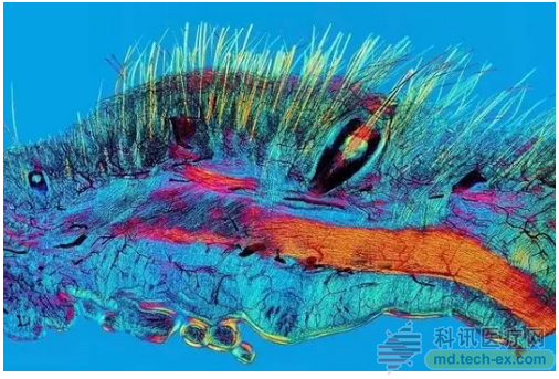
This is the cat's skin under a polarized light microscope that allows only light in a specific direction to pass through to see the cat's hair (yellow), whiskers (also yellow) and blood supply (black).
Iris Clip (Iris Clip)
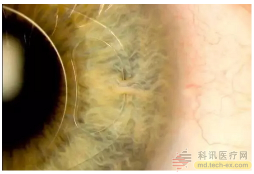
The image shows how to assemble an "iris clip" (also known as an artificial intraocular lens (IOL)) onto the eye. The device is secured to the iris by a small surgical incision for the treatment of myopia and cataracts.
Breastcancer Twitter Connections (association between breast cancer and Twitter)
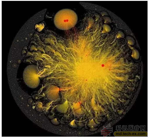
The above image is a graphical representation of the data collected by the tweet containing the label # breastcancer. Twitter users are represented by points (called nodes), and lines connecting nodes represent relationships between users. The size of nodes varies according to the number and importance of other nodes they are connected to. The thickness of each connecting line is determined by the number of times two users interact or are mentioned to each other. The "double yellow" structure at the top of the image (a large yellow circle with two red dots) represents a common hint for both accounts.
MicroRNA Scaffold Cancer Therapy (MicroRNA Stent Cancer Treatment)
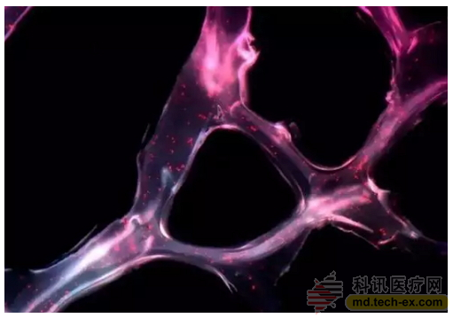
This synthetic network can cover tumors and deliver short gene sequences (called microRNAs) to cancer cells. In this way, the cancer treatment test results in mice showed that the tumor was reduced by 90% in just two weeks.
Hawaiian Bobtail Squid (Hawaii Mackerel)
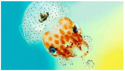
Hawaiian mackerel, which lives in the Pacific Ocean, is a nighttime predator. It is buried under the sand during the day and catches shrimp near the coral reef at night. Underneath these aquatic animals is a light organ with a lot of glowing bacteria called Vibrio fischeri. Squid provides food and shelter for these bacteria, and these bacteria are rewarded with radiance.
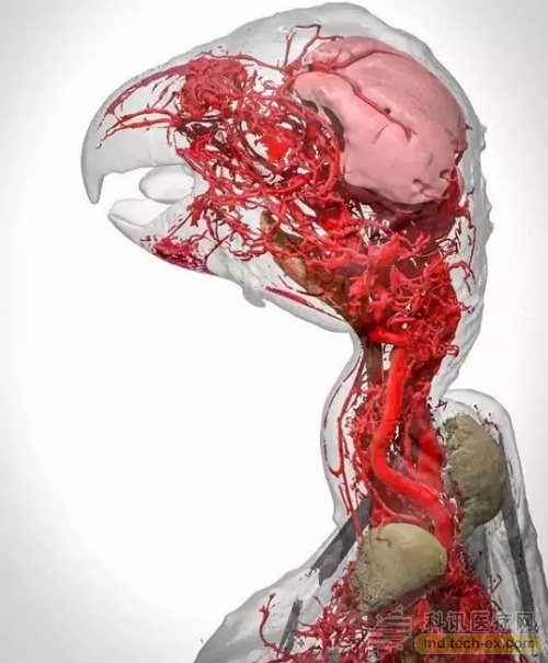
Using CT scanning and digital imaging, the researchers were able to see the entire vascular network of the pigeons, even to the capillary level. At the bottom of the picture, the intricate network of blood vessels in the neck of the pigeon can be seen, and this happens to take care of the blood supply under the skin of the pigeons, allowing it to control its body temperature.
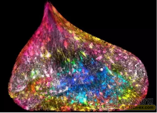
Brain on a Chip (the "brain" growing on plastic sheets)
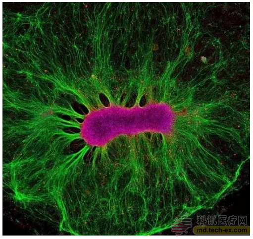
Neural stem cells grow on synthetic gels. Although in vitro, these stem cells (shown in magenta in the figure) are capable of producing nerve fibers (shown in green). Researchers are currently looking for a path to cultivating micro-organs on plastic sheets that eventually become interrelated. These systems can be used to accurately predict the effectiveness and toxicity of drugs and vaccines, and do not require animal testing in medical research.
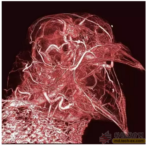
Blood Vessels of an African Grey Parrot
The picture shows the 3D reconstruction of the African Grey Parrot. The model uses a new contrast agent called BriteVu that details the highly complex vascular system in the bird's head and neck. This new contrast agent makes it possible to vividly restore those incredible details that have reached the subject's capillary level.
Through the development of science and technology, we can peep into more mysteries of life. Which of these "live" pictures do you like the most?
Source: 36æ°ª
Freeze Dryer,Food Drugs Lyophilizer,Vacuum Lyophilizer Drying,Chemical Freeze Drying
Guangdong Widinlsa International Co.Ltd , https://www.guangdongwidinlsa.com