Oncology research is one of the hotspots of small animal PET/CT molecular imaging technology, including the following important aspects: establishing monitoring of tumor biological models, screening tumor models, and evaluating the efficacy of new drugs or treatments.
1 Tumor model monitoring In vivo anti-tumor tests during drug development usually require the use of animal tumor transplantation models. Successful tumor transplantation represents the successful establishment of a tumor model. Because PET imaging not only shows the location of the tumor, but also shows the performance and activity of tumor-specific molecules and biological processes, and is a non-invasive method for continuous observation. Therefore, PET/CT is a small animal to evaluate whether the tumor model is An important imaging method has been established for success.
The figure below shows a case in which Super Nova ® PET/CT was used to track and observe the size and growth of tumor models during tumor model establishment.

Establishment of rat liver orthotopic tumor model
(3D correlation shows tumor location; scan for 10 min after injection of 0.4 mCi FDG for 1 h)
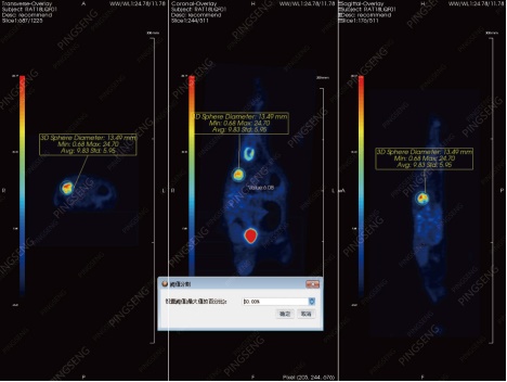
Spherical ROI delineation to obtain tumor
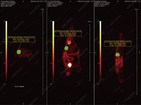
Threshold extraction of high uptake FDG area to obtain tumor volume
2 Tumor model screening Due to the non-invasive imaging characteristics of PET/CT, it can also be used for the screening of tumor model preparation. The following is a high-throughput model screening of a small cell model of small cell lung cancer using a Super Nova® PET/CT (single health care) to observe the effect of modeling and evaluate the tumor model.
2 Tumor model screening Due to the non-invasive imaging characteristics of PET/CT, it can also be used for the screening of tumor model preparation. The following is a high-throughput model screening of a small cell model of small cell lung cancer using a Super Nova® PET/CT (single health care) to observe the effect of modeling and evaluate the tumor model.
Non-small cell lung cancer Zr89 PET/CT imaging
3 Efficacy evaluation In the application of molecular biological models of tumors, it is usually necessary to select new molecular targets to establish corresponding treatment methods according to the characteristics of tumor cells, or to synthesize corresponding targeted drugs for treatment. The evaluation of the efficacy of new drugs or treatments is based on the tumor model. Small animal PET/CT provides an effective observation method for efficacy evaluation and tumor monitoring.
- Example 1:
Published papers:
131 I -Labeled Copper Sulfide-Loaded Microspheres to Treat Hepatic Tumors via Hepatic Artery Embolization
Accept the magazine: Theranostics
Impact factor: 8.766
In this paper [1] , the imaging method of small animal PET/CT was used to analyze the efficacy of the new tumor treatment method. In this study, a multi-functional diagnostic integrated microsphere 131 I-HCuSNPs-MS-PTX was prepared for minimally invasive transcatheter hepatic artery embolization for liver tumors. This compound combines chemotherapy, radiation therapy and photothermal therapy. Multiple characteristics. The analysis results of the Super Nova® PET/CT image are one of the important indicators for evaluating the efficacy of the compound micro-effect method.
- Tumor model establishment
During the study, the researchers used a subcutaneous vaccination method to implant Walker-256 tumors on donor mice and monitor the growth of tumors by scanning with Super Nova® PET/CT. After the tumor reached 6-8 mm, the tumor of the donor mouse was chopped into small pieces of 1 mm 3 and transplanted into the right lobe of the recipient mouse.
- Experimental methods and processes
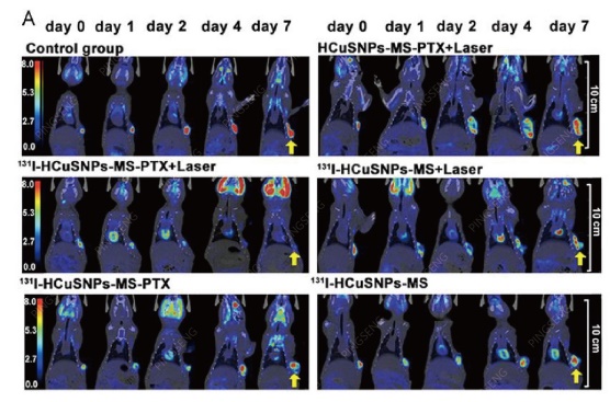
Ten days after the recipient mice received the tumor transplant, the tumor size was monitored by Super Nova® PET/CT (health care), and when the tumor grew to 6-8 mm again, it was treated by injection of microspheres, and before and after treatment 1- 7 days, the small animal PET / CT scan volume observation and SUV value analysis, to obtain the efficacy of minimally invasive treatment of the tumor: experimental group 131 I-HCuSNPs-MS-PTX plus laser treatment on the first day after the tumor pair The absorption of FDG decreased significantly, and there was no FDG absorption after 7 days. In the control group, I-HCuSNPs-MS-PTX plus laser treatment decreased the absorption of FDG in tumors after 1 day, but increased rapidly after 2 days; other treatments were not used, or only 1 was used. The control group of the compound or the compound of the two therapeutic properties showed tumor growth.
- Experimental results
The investigators believe that the multi-functional diagnosis and treatment integrated microspheres 131 I-HCuSNPs-MS-PTX, +Laser for minimally invasive transcatheter hepatic artery embolization for liver tumors have clinical application prospects.
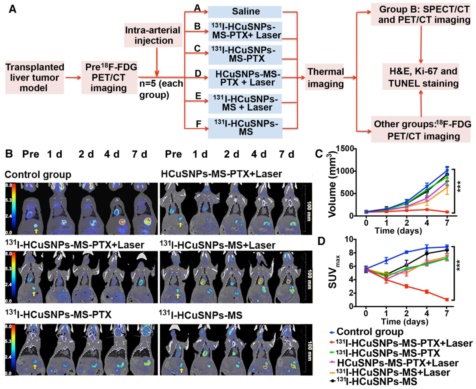
Effect of PET/CT on small animals in different treatments of rat liver tumors
- Example 2:
The combined therapeutic effects of 131 iodine-labeled multifunctional copper sulfide-loaded microspheres in treating breast cancer
Accepted by the journal: Acta Pharmaceutica Sinica B
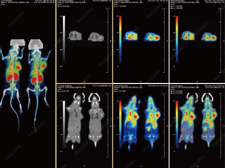
In the same year, the researchers used Super Nova® PET/CT (Health) to observe the efficacy of this drug and treatment in the treatment of breast cancer and published a new paper [2] . The researchers planted breast tumor cells into the second breast fat pad of the rat (female) and continued to observe until the tumor grew to 5-6 mm, followed by injection of microspheres for treatment, while being compared to other treatment modalities. Super Nova® PET/CT was used to follow up the treatment during this evaluation. The results showed that 131 I-HCuSNPs-MS-PTX+Laser was also effective in the treatment of breast tumors.
Accepted by the journal: Acta Pharmaceutica Sinica B
In the same year, the researchers used Super Nova® PET/CT (Health) to observe the efficacy of this drug and treatment in the treatment of breast cancer and published a new paper [2] . The researchers planted breast tumor cells into the second breast fat pad of the rat (female) and continued to observe until the tumor grew to 5-6 mm, followed by injection of microspheres for treatment, while being compared to other treatment modalities. Super Nova® PET/CT was used to follow up the treatment during this evaluation. The results showed that 131 I-HCuSNPs-MS-PTX+Laser was also effective in the treatment of breast tumors.
Rats under the fat pad of breast tumors, PET/CT effect maps of different treatments (three-dimensional correlation showed tumor location; 10 minutes after injection of 0.8mCi FDG for 1 hour)
Summary: Small animal PET/CT has played an increasingly important role in oncology research and is a sharp tool in pre-clinical medical research.
Small animal PET/CT equipment - Super Nova® PET/CT (Ph. Health)
appendix:
In PET/CT oncology studies, radionuclides are commonly used to label drugs or substances involved in the physiological metabolism of organisms, such as 18 F-labeled 18 F-FDG, which are locally radioactive after uptake of 18 F-FDG by tumor tissues with high glucose metabolic rate. The concentration is multiplied, and a strong contrast is formed on the PET image. This contrast is enhanced with the degree of malignancy of the tumor, and the tumor can be monitored and analyzed in the living state of the organism to obtain diagnostic data. The following are other commonly used drugs for PET tumor research:
| drug | Biological process |
| 18 F-FDG | Glucose metabolism |
| 11 C-acetate | Cell Proliferation |
| 11 C-methionine, etc. | Amino acid metabolism |
| 18 F-choline, 11 C-choline | Cell membrane formation |
| 18 FLT | Nucleic acid metabolism |
| 18 F-DOPA | Tumor receptor |
| 18 F-FES | Estrogen |
| 69 Ga-DOTA-TOC | Somatostatin receptor |
| 18 F-FMISO, HX-4, etc. | Hypoxia metabolism |
Note:
[1]. Qiufang Liu, Yuyi Qian, Panli Li, Shaoli Song, et al. 131I-Labeled Copper Sulfide-Loaded Microspheres to Treat Hepatic Tumors via Hepatic Artery Embolization [J]. Theranostics, 2018, 8(3): 785- 799; (IF=8.776)
[2]. Qiufang Liu, Yuyi Qian, Panli Li, Shaoli Song, et al. The combined therapeutic effects of iodine-labeled multifunctional copper sulfide-loaded microspheres in treating breast cancer [J]. Acta Pharmaceutica Sinica B 2018;8(3 ): 371–380
For more case sharing, please pay attention to the public account

Life, born for scientific research <br> Please support domestic manufacturers of high-end scientific research equipment!
Website: E-mail:
Changzhou Ziying Metal Products Co., Ltd , https://www.ziyingmetal.com