Stem cell transplantation has potential clinical application prospects for repairing damaged myocardial tissue. Some researchers have injected stem cells into the myocardium as a biological pacemaker. However, if these cells deviate from the transplant site, local arrhythmia will occur, potentially fatal risk. . Therefore, there is an urgent need for in vivo tracer observation of transplanted stem cells. Green fluorescent protein and traditional fluorescent dyes are interfered with by autofluorescence in the body imaging, while nuclear imaging methods such as MRI and PET have higher requirements on the number of cells.
New York State University Rosen AB uses negatively charged carboxyl quantum dots to label mesenchymal stem cells. In vitro experiments show that quantum dots do not affect stem cell proliferation and differentiation, nor affect the transfection and expression of stem cells. . After transplanting stem cells labeled with quantum dots into the myocardial injury site, the quantum dots maintained good fluorescence brightness and diffuse distribution of cytoplasm for six weeks, and did not enter adjacent cardiomyocytes during stem cell division (Fig. 1). The frozen sections of myocardial tissue after stem cell transplantation for 1h and 1d were used to realize the in vivo three-dimensional reconstruction of transplanted stem cells by quantum dot single-cell imaging technique and related algorithms (Fig. 2). In conclusion, this study clarified the good application of quantum dot labeling in stem cell tracing, and realized the three-dimensional fluorescence imaging of transplanted cells for the first time, which is of great significance for the study of myocardial tissue regeneration.
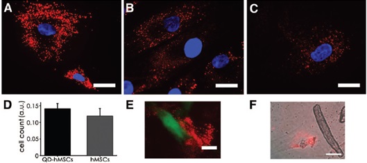
Figure 1 Six weeks after the implantation of quantum dot-labeled mesenchymal stem cells into a damaged heart, the quantum dots remain fluorescent and diffuse in the cytosol.
(A) 2 days; (B) 16 days; (C) 44 days;
(D) The results of mitochondrial lactate dehydrogenase experiments show that quantum dots have no effect on the proliferation of mesenchymal stem cells;
(E) quantum dots (red) do not enter adjacent cardiomyocytes (green);
(F) Cells were cultured in vitro for lysis, and the quantum dots (red) did not enter adjacent cardiomyocytes.
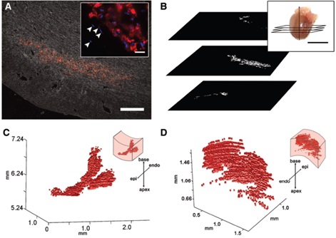
Figure 2 Quantum dot-labeled mesenchymal stem cells were transplanted into a damaged heart, frozen local tissue and sections were prepared, and in-vivo three-dimensional fluorescence imaging was performed using quantum dot single-cell imaging techniques and related algorithms.
(A) quantum dot-labeled mesenchymal cells (red) are distributed in myocardial tissue;
(B) cross-sectioning of frozen myocardial tissue;
(C) Three-dimensional reconstruction of quantum dot-labeled stem cells 1 h after transplantation;
(D) Three-dimensional reconstruction of quantum dot-labeled stem cells 1 day after transplantation.
Source of the document:
Rosen AB, Kelly DJ, Schuldt AJ, Lu J, Potapova IA, Doronin SV, Robichaud KJ, Robinson RB, Rosen MR, Brink PR, Gaudette GR, Cohen IS. Finding fluorescent needles in the cardiac haystack: tracking human mesenchymal stem cells labeled With quantum dots for quantitative in vivo three-dimensional fluorescence analysis. Stem Cells. 2007;25(8):2128-38.
GMP Certificated Immune Globulin Injection Supplier in China
Hepatitis B Immunoglobulin,Hep B Immunoglobulin,Hepatitis B Immunoglobulin Vaccine,Hepatitis Immune Globulin
FOSHAN PHARMA CO., LTD. , https://www.full-pharma.com